What is Kidney Cancer?
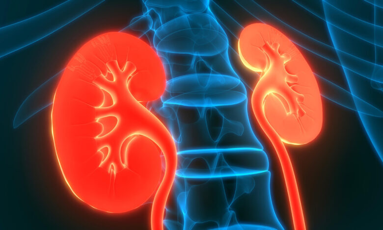
There are two kidney-like kidneys in the human body, one on the right and the other on the left. Protected by the lower part of the rib cage, the kidneys rest against the posterior abdominal wall. In the upper part of the kidneys are the adrenal glands. Both the kidney and the adrenal gland are covered by a thin fibrous membrane structure.
Kidneys have a very important task for our body, such as filtering waste materials. In addition, the secretion of the hormone called renin, the regulation of blood pressure and fluid balance, and the production of the hormone erythropoietin, which provides the formation of red blood cells, are among the functions of the kidneys.
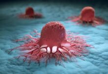
Benign and malignant tumours are also encountered in the kidneys. Cyst development, on the other hand, is a generally seen condition in the kidney, but it is not considered as cancer. In non-life-threatening cysts, only monitoring can usually be performed without the need for treatment. This type of cancer, which is more common in men, can be cured with early diagnosis and then point-and-shoot treatment methods, as in many cancer types.
Symptoms of kidney cancer include haematuria, blood in the urine, low back pain, swelling in the waist, loss of appetite, weakness, and unexplained weight loss.


Causes of Kidney Cancer
Smoking is an effective element in many diseases. It also carries a risk for renal (kidney) cell carcinoma. Increasing smoking will increase the risk of cancer in direct proportion. Kidney cancer mostly seen in older people.
It is more common especially in men over 60 years of age. Cholesterol metabolism and certain changes in the immune system increase the risk in people who are overweight. Doing sports, which is a factor that reduces the risk of cancer, prevents weight gain and keeps the body function in balance. Medications taken to lower high blood pressure are thought to increase the risk of cancer.
However, it is not known exactly whether the changes that occur as a result of high blood pressure or the medications taken pose a risk. In addition, functional weakness due to chronic kidney diseases is thought to increase the risk of cancer. The risk of cancer is considerably increased in people who have had an organ transplant.
It is especially increased in kidney transplantation. It is essential that these people pay more attention to their health and have the necessary health checks done. Environmental factors, such as certain chemicals, are also thought to increase the risk of cancer. However, family genetic factors are also an important factor in cancer risk.
What are the Types of Renal Cell Carcinoma?
The most common kidney cancer is Renal cell carcinoma. In this type, one tumour usually develops in the kidney, but in some cases, two or more tumours can be found. There are many subtypes of this carcinoma and the identification of the species will be helpful in deciding the treatment. In clear cell carcinoma, the cells stand out as very pale or clear.
This type, which is generally seen with venous invasion, metastasizes to the lung, liver and soft tissue most frequently. In papillary renal cell carcinoma, there is a single or multiple layer of cells around the fibro vascular nucleus to form the papillary structure. It usually follows a positive course and is detected at an early stage. Chromophobe renal cell carcinoma, collecting duct carcinoma, and multilocular cystic renal cell carcinoma are also among the subtypes.
Diagnosis in Kidney Cancer
A retrospective analysis is made by listening to the patient’s complaints. After the physical examination, blood and urine tests are requested. The results are examined and tests such as magnetic resonance (MR), computed tomography (CT), ultrasonography and intravenous urography are also requested for the analysis of the kidney and all structures around it.
In the physical examination, it is tried to determine whether there is a hardness or a mass different from the normal in the abdomen (abdominal) area. Then, in the tests requested, the general health status of the person whose disease is suspected is analyzed. The spread of the cancer is also checked. The next step is to make a treatment plan according to the results and analysis.
Blood and Urine Analysis
In these tests, the status of red blood cells is checked with a blood test, and the high number of cells suggests that an excess of erythropoietin hormone is produced by the kidneys. It can also be kidney cancer cells that do this. In addition, the amount of enzymes in the liver and calcium in the blood are also examined. A higher than expected calcium level may suggest that cancerous cells may have spread to the bones.
In the urinalysis, bacteria, cancer cells or bleeding in the urine are analysed. Since most individuals with kidney cancer have bleeding in the urine, this test is very important and is frequently used for diagnosis.
Kidney Cancer Stages
After a patient is diagnosed with kidney cancer, determining the stage and the course of the spread is very important in terms of treatment planning. Accordingly, the appropriate treatment method to be applied is selected. Staging is determined by the spread of cancer to the kidneys and other parts of the body. This situation can also be called the concept of measuring the depth and width of the disease.
In some cases, the stage of the disease can be determined during diagnosis. However, to be sure, additional imaging techniques such as computed tomography (CT), magnetic resonance (MR), intravenous pyelogram, bone scan, angiography or ultrasound may be needed. As a result of all these examinations, individual-specific treatment plans are made, taking into account the extent of the cancer’s spread.
It is very important for treatment planning to know the region where the disease has spread and the degree of aggressiveness in staging. The early stage of the cancer is indicated by the number 0, while the most advanced stage is symbolized by the number IV. The characteristics of the phases are stated as follows.
Kidney Cancer Stage 1
Cancer cells are only found in the kidney and no spread to distant organs or lymph nodes can be seen.
Kidney Cancer Stage II
The tumour is still in the kidney. Cancer cells have not spread to distant organs or lymph nodes. But its size is larger than stage I. Stage 3 in kidney cancer is observed in two ways.
Kidney Cancer Stage IIIa
Cancerous cells have begun to appear in the renal vein and the main vein or in the structures surrounding the kidney around the kidney. There was no spread to the adrenal gland or distant organs. No cancerous cells exist in the lymph nodes.
Kidney Cancer Stage IIIb
Cancerous cells can occur in different sizes outside the kidney. It does not spread beyond the membrane surrounding the kidney. Cancer has spread to surrounding lymph nodes. Cancerous cells are not seen in distant lymph nodes or distant organs. Like the 3rd stage of kidney cancer, the 4th stage is examined in two ways.
Kidney Cancer Stage IVa
The mass occurs beyond the membrane (greto fascia) surrounding the adipose tissue near the kidney. It can also be seen in the adrenal glands. It may spread to nearby lymph nodes. There is no metastasis to distant lymph nodes or organs.
Kidney Cancer Stage IVb
The tumour, which can be of different sizes, may grow outside the kidney. Cancer cells can metastasize by spreading to lymph nodes or organs located away from the kidney.
Imaging Tests
These tests may use magnetic fields, sound waves, x-rays, or radioactive products. These are the tests applied to determine whether the suspected area contains tumour cells, to determine the way of spread, to analyse the effectiveness of the treatment and to understand the traces of recurrence. Unlike many cancers, kidney cancer can usually be diagnosed with imaging tests without a biopsy, but there are cases that require a biopsy.
Computed tomography (CT), ultrasound and magnetic resonance (MR) are helpful methods in the diagnosis of kidney cancer. Apart from that, chest X-ray and bone scan are mechanisms used to test the spread of cancerous cells to different parts of the body.
Computed tomography (CT)
In this three-dimensional imaging, possible abnormalities and examination are performed on the tumour. It gives information about the size and location of the audience. In some cases, detailed information is obtained by administering contrast material.
Magnetic Resonance Imaging (MRI)
Soft tissue images are taken using radio waves and strong magnets. The energy from the radio waves is absorbed and transferred to the suspect area. The computer presents the image in detail as cross-sectional images.
Ultrasound
Internal organs are visualized using sound waves. The echoes of the emitted sound waves coming from the kidney are recorded and transferred to the visual format. It can also help to remove a mass to support the biopsy.
Positron Emission Tomography (PET)
A radioactive isotope called FDG is injected into the individual through the intravenous line and the differences in the tissues are detected. Positron irradiating FDG (fluorodeoxyglucose) is used by cancer cells as a food source, and PET can detect adherent cancer cells. It is essential for both the status of the primary cancer and the control of whether it has spread to other organs.
Intravenous Pyelogram
Contrast material is injected intravenously to the sick person. In this way, images are taken by filling the bladder, kidneys, urinary channels.
Angiography
In this method, which also uses contrast material, a probe is inserted through the leg artery and extended to the artery going to the kidney. By applying dye injection, the vessels feeding the tumor in the kidney are determined and plans are made.
Bone Scan
The low dose and low amount of the substance injected intravenously is localized to the cancerous areas in the bone. The spread to the bones is observed by imaging with a special camera.
Fine-Needle And Coarse-Needle Biopsies
Biopsy is the analysis of the sample obtained from the suspicious area as a result of laboratory tests. Accordingly, biopsy is used for masses that cannot be fully understood by imaging. There are 2 types of biopsy methods in kidney cancer as thick and fine needle biopsy. In both, the needle is inserted through the skin into the area where the tumour is, and a sample is taken. During this process, ultrasound or computed tomography can be used as support to reach the right area.
Fuhrman Rating
In this method, the mass taken by biopsy is analysed with a microscope. With this grading, information about the course of the cancer is obtained.
How Is Kidney Cancer Treated?
In treatment, surgical methods, radiotherapy, chemotherapy, ablation, smart drugs and immunotherapy methods can be applied according to the individual’s condition.
Kidney Cancer Surgery
When cancerous cells are found outside of the kidney, the whole or cancerous part of the kidney can be surgically removed and treated. However, if the cancer has spread to different areas, the surgical method is not a complete solution, but it can reduce the symptoms of the individual. In partial nephrectomy, the cancerous area is removed. The entire kidney is removed in radical nephrectomy.
Chemotherapy
It is the administration of drugs to the body to kill cancerous cells. Oral or intravenous injection is used. These given drugs travel throughout the body through the circulation and are effective in the treatment of tumours that have spread throughout the body. However, chemotherapy usually shows resistance in kidney cancer cells. There are few chemotherapy drugs that benefit patients with kidney cancer.
Radiotherapy
It aims to kill cancerous cells by sending high radiation energy. It can be applied externally as radiotherapy. Radiation is sent from outside the body to the area where the kidney is located. Kidney cancer is generally not very sensitive to radiation. For this reason, it is a method that can be applied to prevent the rapid course of cancer, to control metastases or to keep pain at a minimal level.
Targeted Therapies – Smart Drugs
These treatments are made with anticancer drugs. Some targeted therapies block the growth signal from reaching cancerous cells. Others reduce the nutrition of cancerous cells through the blood. There are also treatments that strengthen the immune system. In this way, the spread of cancer slows down and the defense system becomes stronger and fights cancerous cells more effectively. Smart drugs can be used in combination with other treatment methods.
Immunotherapy Treatment In Kidney Cancer
The aim of immunotherapy is to fight cancer cells and kill cells by making the immune system, which is the defence mechanism in the body, stronger. Today, there are many studies on immunotherapy. New generation immunotherapy drugs not only improve the quality of life of patients with advanced kidney cancer, but also significantly extend the life span. The general working system of these drugs is on the easier recognition and destruction of cancerous cells. Common side effects include fatigue, nausea, difficulty in breathing, diarrhoea and constipation. Cytokines are a group of proteins that allow cells to communicate with each other. The cytokines used in kidney cancer are generally interleukin-2 (IL-2) and interferon alpha. Both can contribute to a certain shrinkage of the tumour.
Heat – Hyperthermia Treatment In Kidney Cancer
Hyperthermia, also called heat therapy, is a treatment applied using high heat. The human body is exposed to a temperature of approximately 39-44 °C. It is a complementary cancer treatment method. It has been observed that this method causes minimal damage to healthy tissues and kills or causes great damage to cancerous cells. Heat therapy can contribute to the shrinkage of the mass by destroying the cancerous cells and damaging the proteins in the cells. There are few studies on the application of this treatment method.